A surgical breast biopsy needs a cut (incision) in your skin. This allows your healthcare provider to take a large piece of tissue from the breast. In fact, sometimes the whole lump or abnormal area is removed. When this is done, it's called an excisional biopsy. In some cases, only a piece of a large tumor or abnormal area may be taken out. This is called an incisional biopsy. The tissue that's removed for both types of biopsies is sent to a lab for testing.
Most people don't need a surgical biopsy. Changed breast tissue can often be removed with a needle biopsy and sent for testing. You mayneed a surgical biopsy if the results from a needle biopsy aren't clear. A biopsy is the only way to know for sure that a change in your breast is breast cancer.
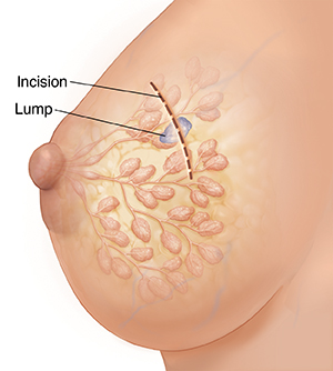
Open surgical biopsy
Open surgical biopsy (incisional or excisional) removes a tissue sample through a small cut in the skin over the lump or abnormal area.
To keep you from feeling pain during the biopsy, you may be given IV (intravenous) sedation and local anesthesia. This means medicines are used to make you sleepy and to numb your breast so you don't feel the surgery. Another choice is general anesthesia. In this case, medicines are used to put you into a deep sleep so you don't feel pain.
Your surgeon then makes an incision in the skin over your breast. If possible, this is done in a way that hides the scar. In most cases, all of the lump is removed along with a margin (edge) of healthy tissue around it. The incision is then closed with stitches. Some stitches dissolve on their own. Others may need to be removed when the incision heals. Your healthcare team will tell you what to expect and how to care for your incision.
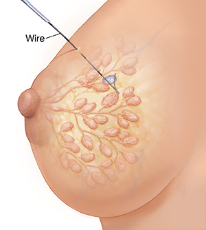
Wire localization
This procedure is needed when a lump in your breast cannot be felt by your healthcare provider. In such a case, a mammogram or ultrasound can be used to find the lump or changed breast tissue before the biopsy. The images are used to guide a thin needle into the area. A thin wire with a tiny hook on the end is put in through the needle. The hooked end is in the area to be removed.
You are then taken to the operating room for surgery. The wire guides the surgeon to the tissue that's removed during the biopsy. It's removed along with the tumor or changed breast tissue.
Featured in